NEW DELHI: In a baffling and exceedingly rare medical case, a 26-year-old man in New Delhi who visited a hospital seeking answers for infertility was shocked to discover a small uterus and fallopian-tube-like structures inside his body.
The bizarre medical discovery was made at RG Hospitals in Rajouri Garden. The patient had sought medical evaluation after failing to father a child. Initial clinical assessments revealed azoospermia—a complete absence of sperm in his semen—along with undescended testicles that remained lodged inside his abdomen rather than settling into the scrotum.
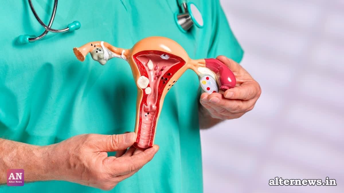
Prompted by these anomalies, doctors ordered an advanced MRI scan, which produced the first major surprise: a small, uterus-like structure visible in the pelvis alongside structures resembling fallopian tubes. Genetic testing confirmed a normal male 46,XY chromosome pattern, pointing medical experts toward a rare congenital condition known as Persistent Müllerian Duct Syndrome (PMDS).
What is PMDS?
Persistent Müllerian Duct Syndrome is an exceptionally rare condition where remnants of the Müllerian ducts—structures that typically develop into the female reproductive tract—fail to regress during embryonic development in individuals with normal male chromosomes and anatomy. Fewer than 300 cases have ever been documented in medical literature globally. While the condition is usually diagnosed during childhood while investigating undescended testicles, discovering it for the first time in a 26-year-old adult during an infertility workup made this case remarkably unusual.
Surgery and Further Shocks
Led by Dr. Susheel Kharbanda, Chief Urologist at RG Hospitals, a multidisciplinary surgical team performed a laparoscopic procedure to investigate further. Inside the abdomen, doctors found a small, underdeveloped uterus, tubular structures resembling fallopian tubes, and severely damaged, atrophied testicles.
Because the undescended testicles carried a high risk of malignancy over time, the surgical team removed both the abnormal Müllerian structures and the testicles.
The biggest shock arrived post-surgery. When pathologists examined the extracted tissue under a microscope, they discovered that the left testicle harbored Germ Cell Neoplasia In Situ (GCNIS)—a pre-cancerous lesion capable of developing into testicular cancer. Blood tests prior to surgery had shown completely normal tumour markers, making the microscopic detection crucial.
"This was an extremely unusual case because the patient came to us with infertility, and the underlying condition had remained undiagnosed until adulthood," said Dr. Kharbanda. "We had to approach the surgery carefully, taking into account both the unusual anatomy and the long-term health risks."
The patient was successfully managed by a coordinated team of specialists spanning urology, radiology, pathology, reproductive medicine, and endocrinology, turning what started as a routine fertility check into a life-saving medical intervention.